MutaTrace — Tracing a tumour to its source
Computational Oncology · Case Study

MutaTrace

Tracing a tumour to its source by reading its mutational fingerprint.

When a cancer is found but its origin is not, treatment stalls. MutaTrace infers a tumour's tissue of origin from its somatic mutations alone — no imaging, no histology — at 90% accuracy across breast, lung, and colorectal cancer.

C>AC>GC>TT>AT>CT>G
Live SBS-96 spectrum — the real mutational fingerprint of breast carcinoma (TCGA, n≈8,000 substitutions)
90.0%
Tissue-of-origin accuracy
2,098
Real TCGA patients
450k
Somatic mutations
3
Tissues · extensible to 33
The problem
In 3–5% of metastatic cancers, the tumour is found but its primary site cannot be identified.

This is Cancer of Unknown Primary (CUP). Because nearly every therapy is chosen by tissue of origin, these patients are treated half-blind — and CUP carries among the worst outcomes in oncology. The one datum always in hand is the tumour's DNA. MutaTrace turns that DNA into an answer.

The input data

Real tumours. Public data. Every figure traces to a mutation call.

The substrate is the TCGA PanCancer Atlas — de-identified whole-exome somatic mutations from real patient tumours, pulled live from the cBioPortal public API. Nothing here is simulated.

SourceTCGA PanCancer Atlas 2018, via the cBioPortal public REST API
AssayWhole-exome somatic mutation calls (MAF-format variants)
BuildGRCh37 — trinucleotide context fetched from Ensembl
Scale2,098 patients · 450,107 somatic mutations
Breast · 1,009 Lung · 561 Colorectal · 528
One input record · a single somatic mutation
patientTCGA-05-4244 geneKRAS locuschr12:25,398,285 changeC>A  (SNP) effectMissense proteinG12C labelLung · LUAD
≈ 215 such records per patient → one 187-feature profile
cBioPortal API features · gene matrix / TMB / SBS-96 DNABERT context XGBoost SHAP + GPT-4o

Every figure on this page is produced by a numbered script in the public repo, from that one API — reproducible end-to-end with python scripts/01…08.

The method — three layers of forensic evidence

Every cancer records its own history in its genome.

MutaTrace reads that record on three independent axes, then fuses them into one prediction per patient.

Evidence I · what broke

Driver architecture

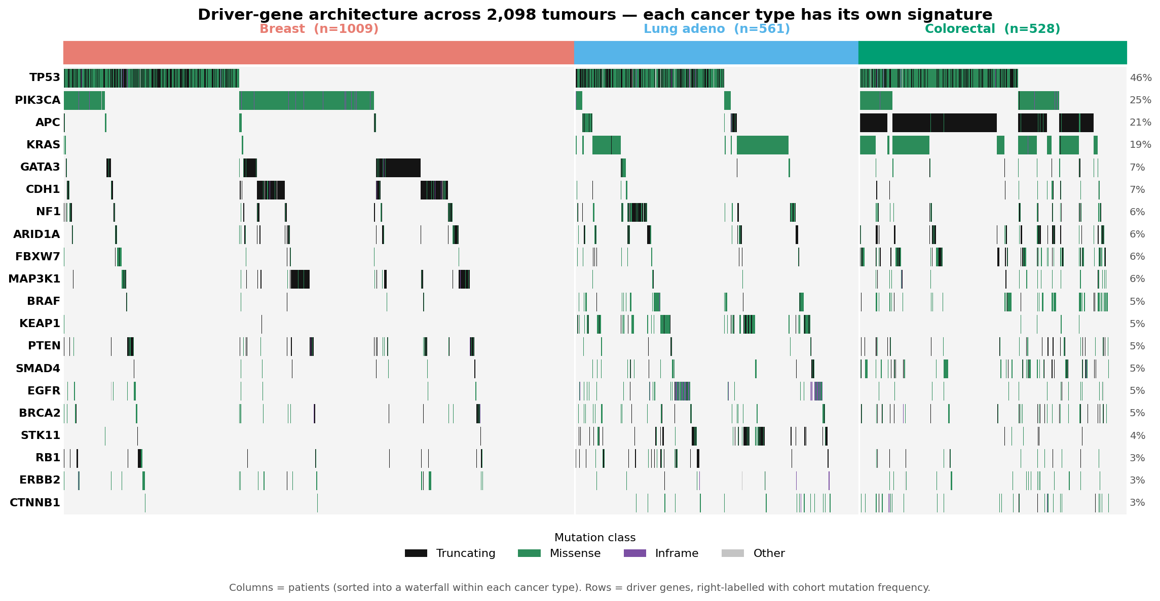
Which cancer genes are mutated. Each tissue carries its own driver signature — the strongest single discriminator.

APC → colonPIK3CA → breastEGFR → lung
Evidence II · what caused it

Mutational signatures

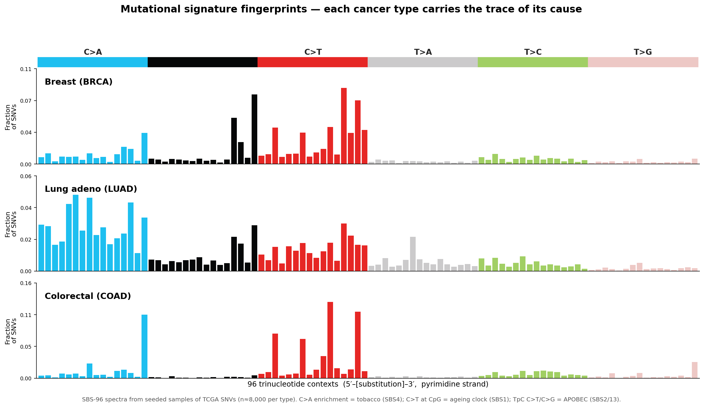
The trinucleotide context of every point mutation encodes its cause — tobacco, APOBEC, an ageing clock — as the SBS-96 barcode.

C>A = tobaccoCpG C>T = ageing
Evidence III · sequence context

Genomic foundation model

DNABERT encodes 129 bp around each driver mutation, capturing local sequence grammar that gene names and signatures miss.

DNABERT · 768-dPCA → 20
The evidence
Plate 01 · Driver architecture Oncoprint of driver-gene mutations across 2,098 tumours
Oncoprint, 2,098 tumours. Each column a patient, each row a driver gene. The black APC block in colorectal, PIK3CA in breast, and EGFR/STK11 in lung are the tissue signatures the classifier exploits. TTN/MUC16 deliberately excluded as long-gene artefacts.
Plate 02 · Mutational fingerprint SBS-96 mutational signature spectra per cancer type
SBS-96 spectra. Lung's towering C>A block is the tobacco signature (SBS4); colorectal's C>T peaks are the ageing/CpG clock (SBS1); breast carries an APOBEC pattern. Trinucleotide context fetched live from Ensembl GRCh37.
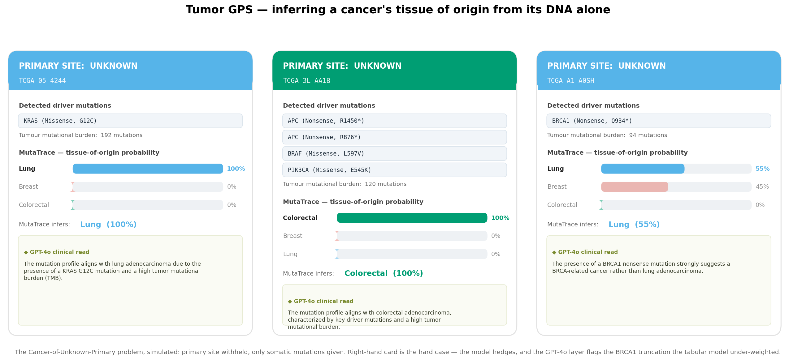
Plate 03 · Tumor GPS Tumor GPS cards inferring tissue of origin for three patients
The CUP inference, simulated. Primary site withheld; only mutations given. Two confident calls, and one honest hard case (right) — the model hedges 55/45 and narrowly misses, while the GPT-4o layer flags the BRCA1 truncation it under-weighted. Probabilities are leak-free (out-of-fold).
The result

90% accuracy, from mutations alone.

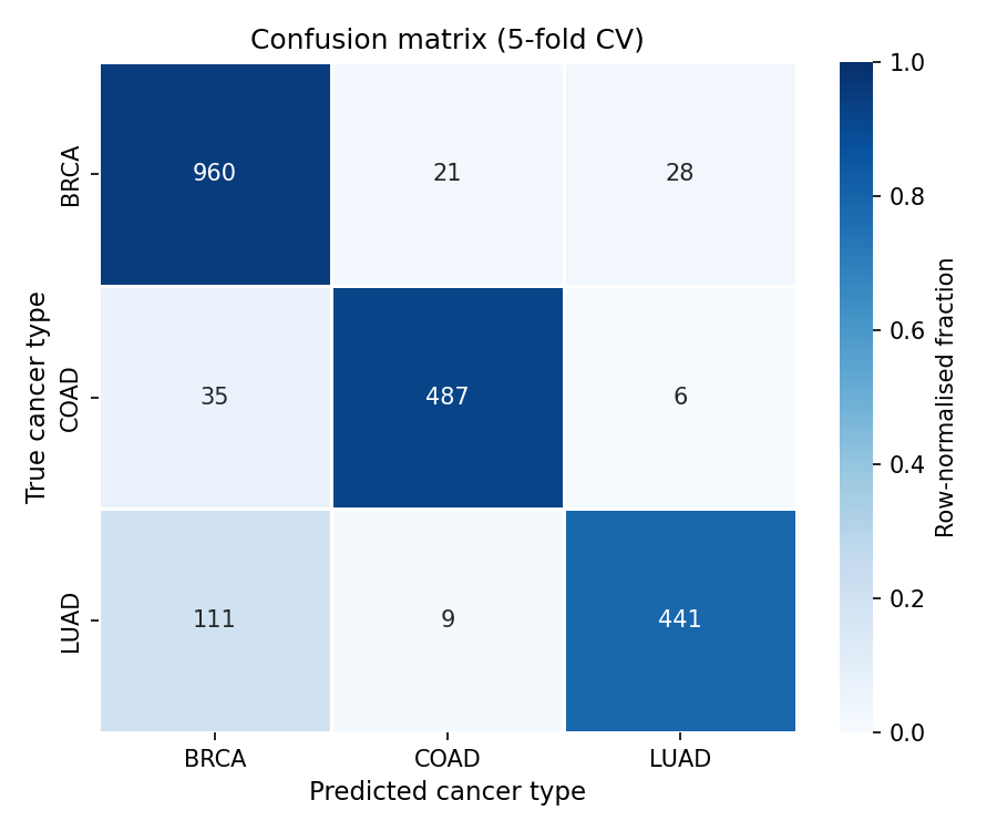
Five-fold stratified cross-validation on 2,098 patients. The signal in somatic mutations is strong enough to place a tumour to its tissue without any clinical, imaging, or expression data.
90.0%
overall tissue-of-origin accuracy · macro-F1 89.7%
random baseline 33% · majority-class baseline 48%
Breast · BRCA
0.91
P 0.87 · R 0.95 · n=1009
Lung · LUAD
0.85
P 0.93 · R 0.79 · n=561
Colorectal · COAD
0.93
P 0.94 · R 0.92 · n=528 — the cleanest call, driven by APC truncation + high burden
Confusion · 5-fold CV Confusion matrix of tissue-of-origin predictions
Lung's lower recall traces to its heterogeneity — EGFR-mutant, KRAS-mutant, and driver-wildtype subgroups, the last of which is genuinely hard from mutations alone.
Built with
DatacBioPortal API · TCGA PanCancer Atlas
SequenceEnsembl GRCh37 REST
Foundation modelDNABERT (6-mer, 768-d)
ClassifierXGBoost · 300 trees
ExplainabilitySHAP · UMAP
ReasoningGPT-4o · LangGraph agent
Features187 · gene matrix + TMB + SBS + DNA-PCs
Validation5-fold stratified CV
Rigour & honest limits

What makes it trustworthy — and where it stops.

Cancer tissue-of-origin classification is a well-established problem with a body of prior work. MutaTrace makes no novelty claim — its value is a clean, interpretable, fully reproducible end-to-end build on public data, not a new method.

No leakage in the demo. Tumor GPS probabilities are out-of-fold — each patient scored by a model that never trained on them.

Curated drivers. TTN and MUC16 excluded — long genes that masquerade as drivers by mutation count.

Interpretable by construction. SHAP attributes every call to genes and signatures; GPT-4o writes the clinical rationale.

Real patient tumours. TCGA PanCancer Atlas, de-identified and public — not simulated data.

Three tissues, not thirty-three. A proof of concept; the architecture is class-agnostic and extends to the full TCGA panel.

Mutations only. Adding copy-number, expression, and methylation — and an independent hold-out cohort — is the path to clinical grade.